
Researchers from the U.S. National Institutes of Health have collaborated with NVIDIA experts on an AI-accelerated method to monitor COVID-19 disease severity from patient chest CT scans.
Published today, the paper studied the progression of lung opacities in chest CT images of COVID patients and extracted insights about the temporal relationships between CT features and lab measurements. A better understanding of the progression of lung opacities in COVID patients could help inform clinical decisions in patients with pneumonia and yield insights during clinical trials for therapies to treat the virus.
Тhe researchers used an NVIDIA Clara AI segmentation model to automate the time-consuming task of segmenting the total lung in each CT scan. Expert radiologists reviewed the total lung segmentation and manually segmented the lung opacities.
To track the progression of the disease, the researchers used generalized temporal curves, which correlated the CT imaging data with lab measurements. Then they used 3D visualizations to reconstruct the evolution of COVID opacities in one of the patients.
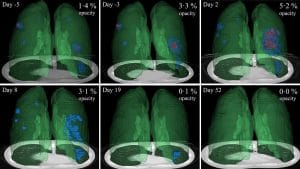
Photo Credits: NVIDIA Developer, News Center
What they found was that lung opacities appeared between one and five days before symptom onset, and peaked a day after symptoms began. They also analyzed two opacity subtypes — ground-glass opacity and consolidation — the ground glass opacities appeared earlier in the disease and persisted for a time after the resolution of the consolidation.
In the paper, the researchers showed how CT dynamic curves could be used as a clinical reference tool for mild COVID-19 cases and might help spot cases that grow more severe over time. These curves could also assist clinicians in identifying chronic lung effects.
The deep learning models were developed using the NVIDIA Clara application framework for medical imaging.